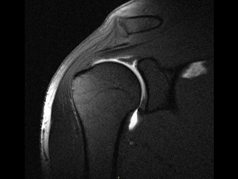
MRI for Joint Analysis
Arthrograms are a series of images of a joint after injection of a contrast medium, usually done by fluoroscopy or MRI. The difference between an MRI with Contrast and an Arthrogram- when an "MRI with contrast" is ordered, contrast is injected into the vein, while the Arthrogram injects contrast directly into the joint under fluoroscopy guidance.
An arthrogram provides a clear image of the soft tissue in the joint (e.g. ligaments and cartilage) so that a more accurate diagnosis about an injury or cause of a symptom, such as joint pain or swelling, can be made. While a plain MRI can provide some information about the soft tissue structures, an arthrogram can sometimes provide much more detailed information about what is wrong within the joint.

