MRI for Spinal Analysis
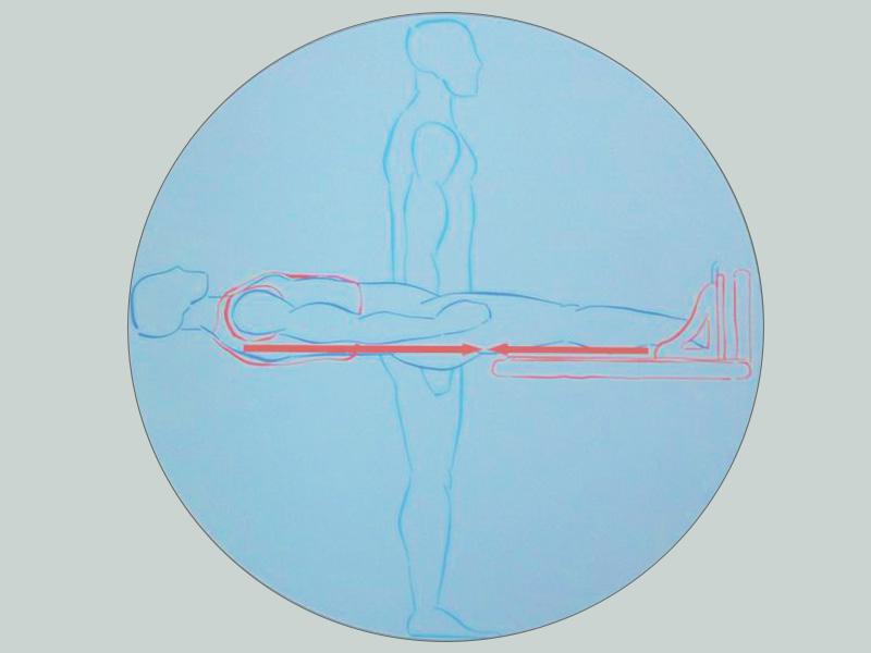
A Weight Bearing MRI, also known as an Axial-loaded MRI, is an imaging test designed to simulate a standing up position that could reveal critical issues not normally visible under a typical MRI. The weight bearing MRI works by applying compression to the lumbar spine and capturing how the patient physically feels while standing. This test could reveal herniation and bulges around the spinal canal not normally visible in a laying-down position.
The simulated weight compresses the spine as if the patient was standing which can reveal herniation and bulges that may not be noticed if the patient was in a relaxed position. This allows a physician more insight into the patient's condition due to higher quality imaging over a standard MRI.

Benefits of a Weight Bearing MRI
The weight bearing technique was developed in 1997 for use in assessing changes of tissue surrounding the spinal canal. It provides a level of image detailing not typically present in normal magnetic resonance imaging, thanks to its visualization of the spine under axial load. If you are experiencing severe or chronic back pain, the issue may only appear while standing, making a normal MRI ineffective. The dynamic nature of the weight bearing MRI allows for positional adjustments to see how rotation, extension, or side-bending can affect your symptoms.
